Research Days 2021 - Artwork
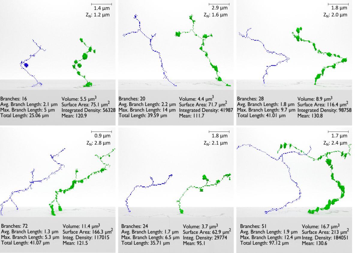
Three-Dimensional Reconstruction and Quantification of PGP9.5-Immunoreactive, Sensory Intra-Epidermal Nerve Fibers
These six 3D renderings were created from confocal imaging of tissue-cleared rat epidermis, ImageJ script automated segmentation, and rendered (visualized) in Blender. This technique provides the ability to measure 3D IENF swelling volumetrically, surface area, total length, individual branch lengths, number of branches, and mean-grey-intensity or integrated density of multiple co-localized markers from immunoreactive (IR) fluorescence. This developing technique allows for IENF quantification in 3D and shows the potential to become a powerful tool for researchers and clinicians. This research was published in Microscopy and Microanalysis on August 1st, 2018, by the Cambridge University Press (https://doi.org/10.1017/S1431927618007274).
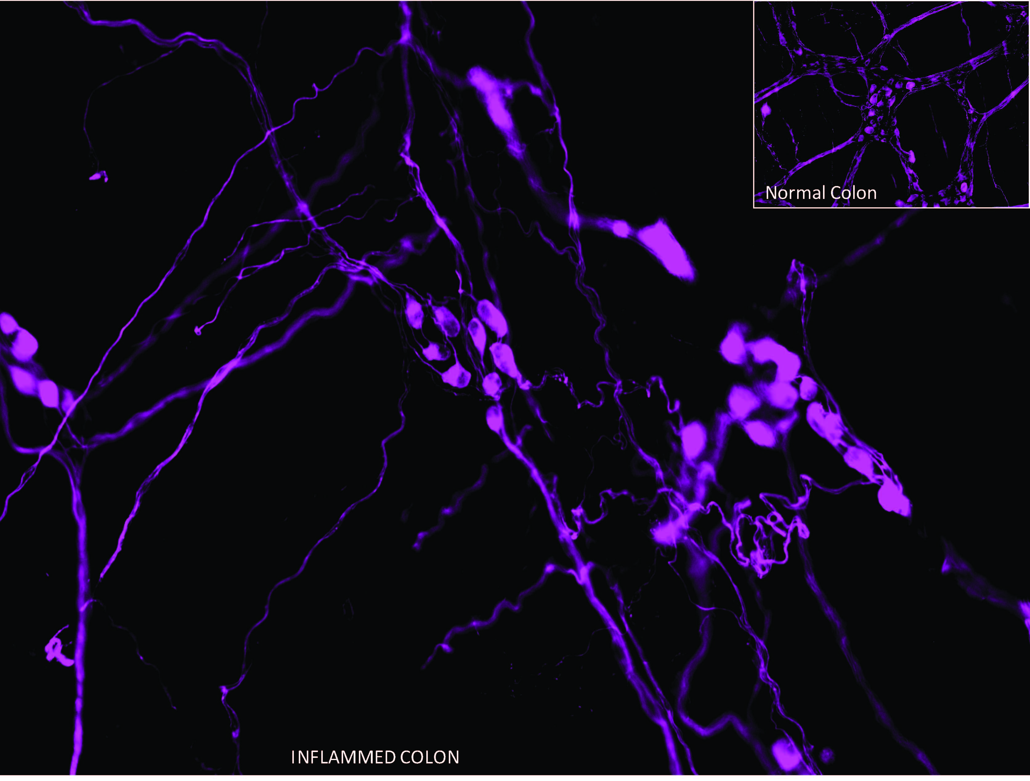
The (GI) Matrix
The myenteric ganglia in enteric nervous system of rat colon. The neurons are labeled with neuronal marker, peripherin. The image was captured and acquired on BX51 epifluorescence microscope (Olympus) at 400x magnification. The image depicts disruption of myenteric ganglia and reduction in number of neurons per ganglia in colon inflammation compared to normal colon (inset).
Canvas 1
Canvas 2
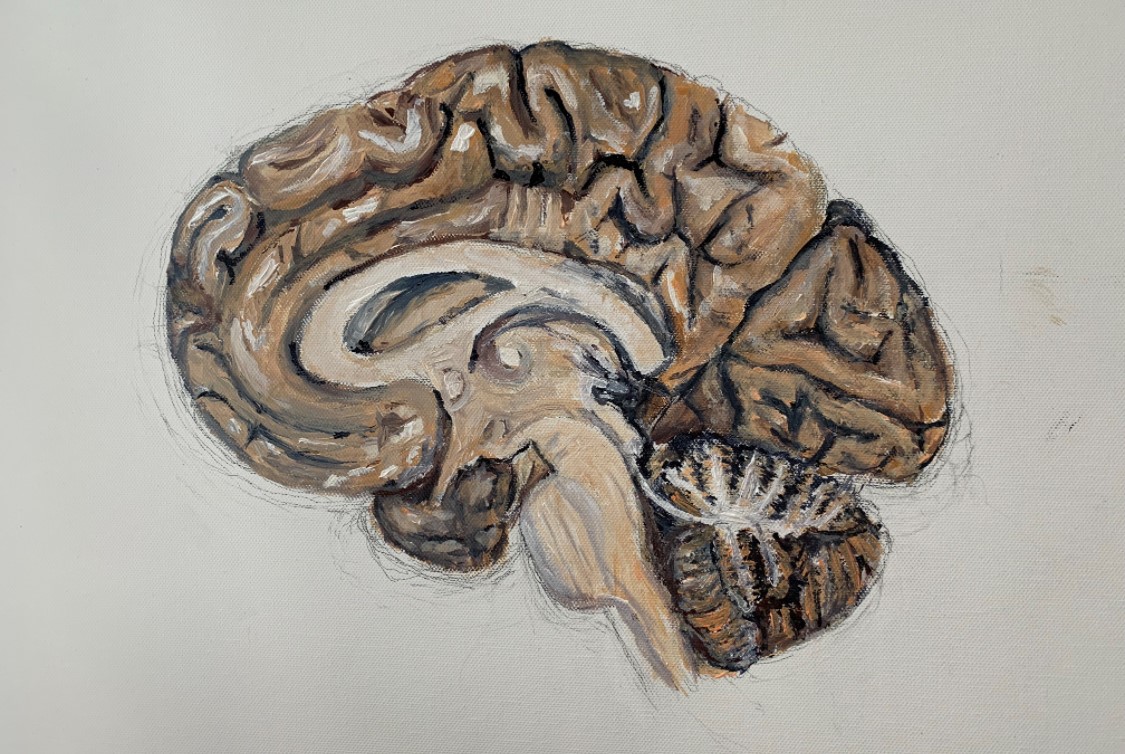
Canvas 3
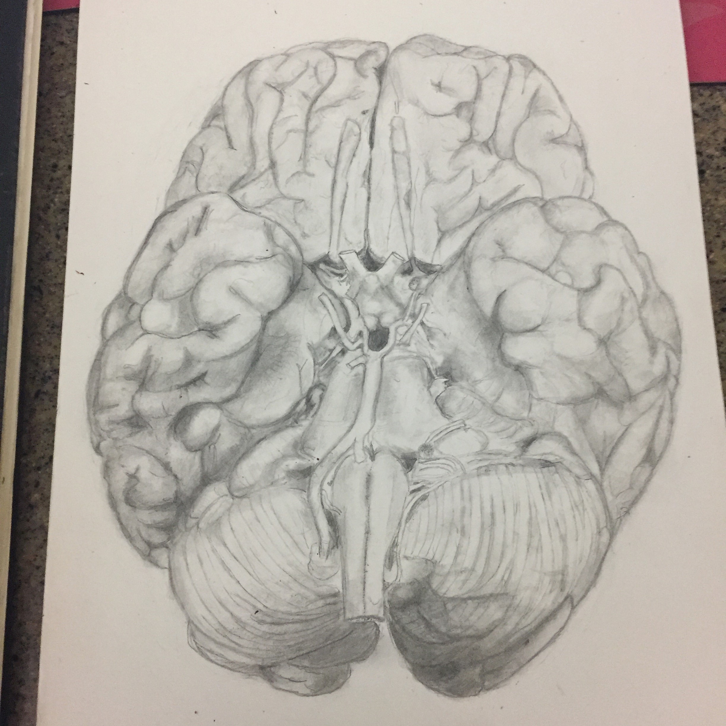
Paper 1
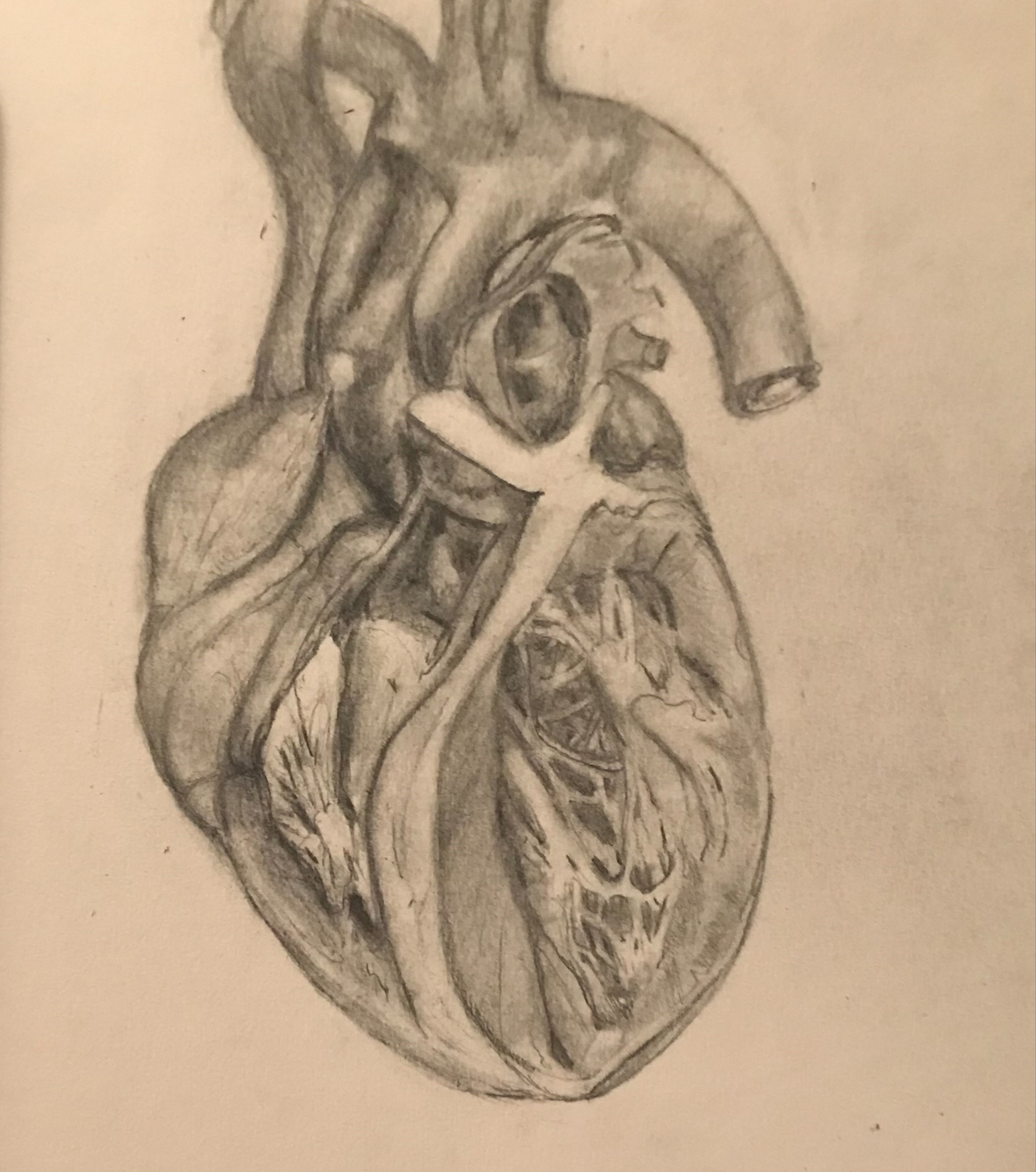
Paper 2
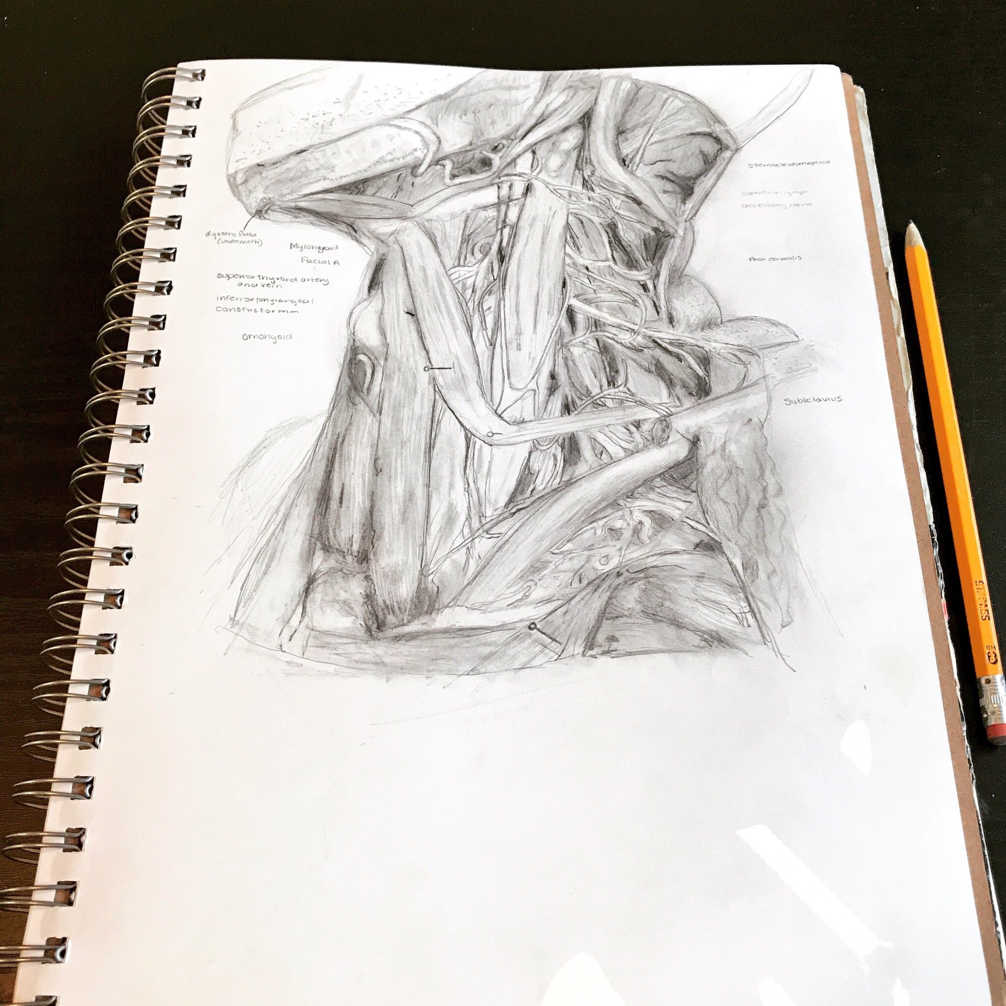
Paper 3
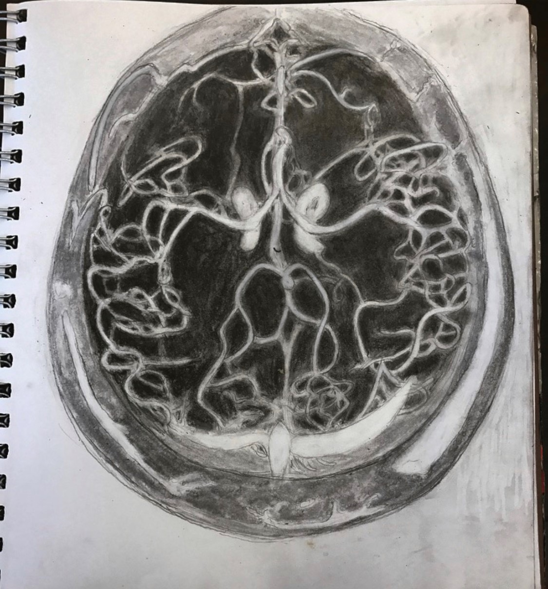
Paper 4
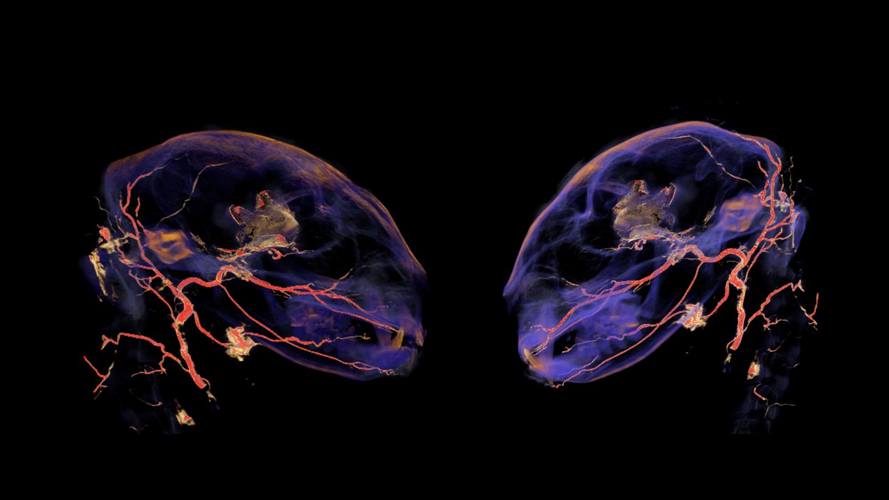
Cranial vasculature of Felis catus
This image depicts a high-resolution CT scan of a Felis catus skull that has had its arterial tree injected with a radiopaque vascular injection medium. The cranial arteries have been isolated and highlighted with Avizo software. A screenshot of the image was taken also using Avizo. The image is part of a larger dataset of carnivoran skulls that have been examined to determine the presence and structure of the carotid rete, a vascular meshwork important to carnivoran thermoregulation.
